
Rock Climbing Finger Pathologies
Scroll down to learn more about our medical illustrations focusing on thumb and long finger pathologies in rock climbing.
Client: Institut für Radiologie und Neuroradiologie
The client, an orthopedic surgeon and climber, approached us to create a series of medical illustrations that explore the intricate ligamentous and tendons structures of the fingers as related to climbing.
01 - Overview
Artists:
Tiffany Fung
Margot Ceelen

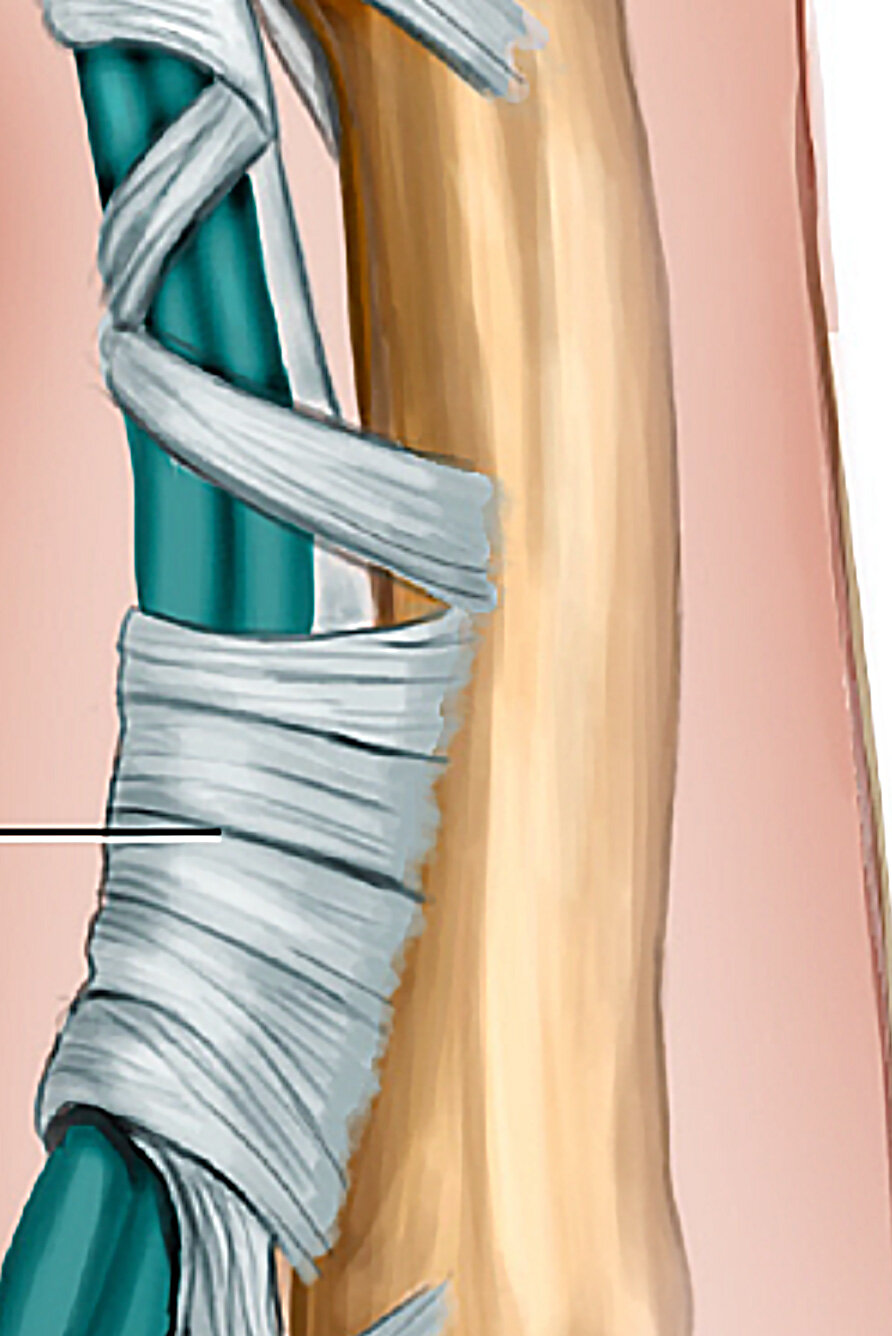
Lateral Finger Anatomy
This lateral view allows you to view the pulley attachments that keep the FDP and FDS close to the bone. In addition, it shows the layered X shape that the collateral ligaments on the joint form.
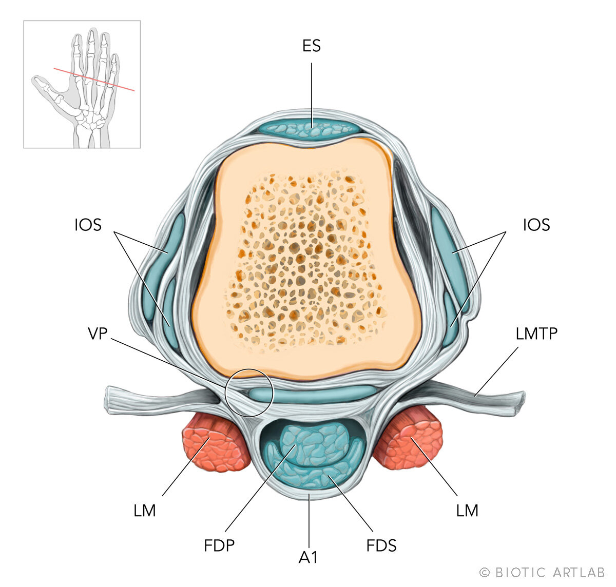
MCP Joint Cross Section
A view of the internal structures at the metacarpophalangeal joint.

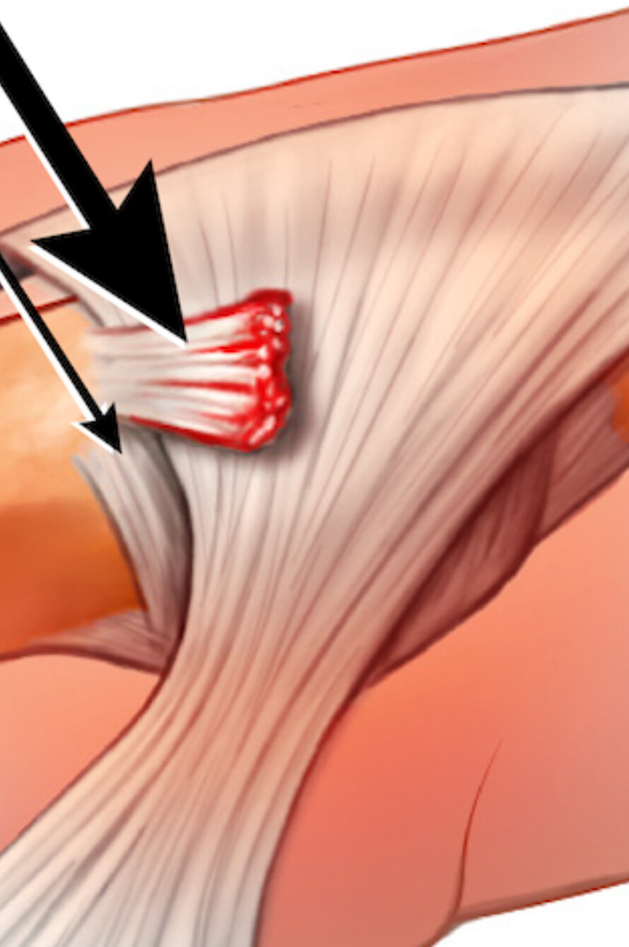
Thumb Stener Lesion
A stener lesion occurs can occur when there is a traumatic injury to the thumb. It occurs when the ulnar collateral ligament (UCL) of the thumb is torn and then becomes stuck on top of the aponeurosis of the adductor pollicis muscle. It is unable to heal naturally and this requires surgical intervention.
02 - What is sports medicine?
Sports medicine is a medical branch that handles physical fitness/performance as well as the treatment and prevention of sports related injuries. The medical experts involved in sports medicine often work together as a team. These team members can include orthopedic surgeons, sports physical therapists/physiotherapists, certified athletic trainers, physical medicine and rehabilitation specialists and SEM (Sports and Exercise Medicine) physicians.
In particular, orthopedic surgeons are focused on the prevention, diagnosis and treatment of disorders of the muscles, joints, ligaments, tendons, and bones.


03 - Our Solution ✨
In order to illustrate the correct anatomy of the hands, we sourced MRI scans of the lateral long finger. From this we were able to extrapolate the structures at the MCP joint. In additions, we show the lateral long finger in an open hand grip to better represent how the pulleys and tendons interact.
Let’s Chat
Do you need medical illustrations to accompany your research in your next research article? Reach out to us and we can start the conversation.